 |  | Marta Campbell |
 |
05.05.2023 11:00 Uhr |
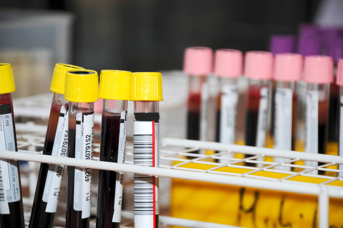
In a small blood count, the doctor counts the platelets (PLT or THRO), erythrocytes (RBC or ERY) and leucocytes (WBC or LEUK). Platelets, also known as thrombocytes, play an important role in blood clotting. / Foto: Adobe Stock/littlebell
A haemogram or haematogram is exclusively concerned with the solid components of the blood, i.e. the red and white blood cells and the platelets. If you want to know whether there are nutrient deficiencies, whether cholesterol or inflammation markers are elevated or what the state of the hormones is, the blood plasma must be examined. Electrolytes, plasma proteins, nutrients, metabolic degradation products and hormones are dissolved in the blood plasma.
In a small blood count, the doctor counts the platelets (PLT or THRO), erythrocytes (RBC or ERY) and leucocytes (WBC or LEUK). Platelets, also known as thrombocytes, play an important role in blood clotting. When a blood vessel is injured, they assemble into plugs to close the defect (platelet adhesion and aggregation). In the process, they release clotting factors and procoagulant messengers. Platelets do not have a nucleus and cannot divide. They develop from megakaryocytes in the bone marrow. If there is a deficiency (thrombocytopenia), there is a risk of bleeding; if there is an excess (thrombocytosis), there is an increased risk of thrombosis.
For a complete blood count, the laboratory determines the haemoglobin (Hb) and haematocrit (HCT or HKT) values. The Hb value indicates the level of red blood pigment. Values that are too low indicate anaemia. The haematocrit value is the percentage of blood cells in relation to the total blood volume. The haematocrit value depends mainly on the erythrocyte count, as red blood cells make up the largest proportion of blood cells. If HCT is too high, there may be an excessive proliferation of red cells or dehydration. Patients with anaemia, blood loss and overhydration have levels that are too low.
Around 99 per cent of blood cells are red blood cells. They are responsible for transporting oxygen in the body. The erythrocytes look like dented discs, have no cell nucleus and cannot divide. The bone marrow produces about 200 billion new red blood cells every day, whose lifespan is 120 days. In order for the body to produce enough erythrocytes, it needs iron. The trace element is a building block for the haem group of the blood pigment haemoglobin. The oxygen and carbon dioxide molecules that transport the red blood cells from the lungs to the tissue or from the tissue to the lungs bind to the iron ion in the centre of the haemoglobin. The number of red blood cells is altered in disorders of the water balance, blood formation disorders and increased new formation (polyglobulia) as a result of a lack of oxygen. The latter is the case, for example, during a stay in the high mountains.
As part of a small blood count, the so-called erythrocyte indices are calculated. They are important to differentiate between different types of anaemia. MCV (mean corpuscular volume) indicates the average volume of an erythrocyte. The value results from the quotient of haematocrit and red cell count. MCH (mean corpuscular haemoglobin, HbE) stands for the average amount of haemoglobin in an erythrocyte. For this value, the haemoglobin value is divided by the erythrocyte count. The mean corpuscular haemoglobin concentration (MCHC) indicates the proportion of haemoglobin in the total volume of red blood cells and is calculated from the haemoglobin/haematocrit quotient or MCH/MCV. An increased red cell distribution width (RDW) is an early indicator of iron deficiency anaemia. Reticulocytes are the precursor cells of erythrocytes. Increased reticulocytes occur with increased new blood formation.
The large blood count consists of the small blood count and the differential blood count. In it, the leukocytes are broken down into their subgroups. Leukocytes are called white blood cells because they have no blood pigment. As part of the immune system, they play a role in both specific and non-specific immune defence. They are involved in infections, inflammations, allergic reactions and autoimmune diseases. Leukocytes make up only 1 per cent of blood cells. When their number is decreased, it is called leukopenia; when it is increased, it is called leukocytosis. Changes in leukocytes indicate a disturbed or activated immune defence. Leukocytes include granulocytes, monocytes and lymphocytes. In order to determine the respective proportions, a blood smear is stained. The percentages are determined from 100 counted leucocytes. The granulocytes are divided into the subgroups neutrophils, eosinophils and basophils depending on the staining of their granules.
Up to 45 percent of leukocytes are lymphocytes. They split into T-lymphocytes and B-lymphocytes. T-lymphocytes become T-helper cells and cytotoxic T-cells after they have had antigen contact. T-helper cells stimulate the immune defence, cytotoxic T-cells destroy foreign and tumour cells. B-lymphocytes develop into antibody-producing plasma cells after antigen contact. The lymphocyte count is increased, as is the eosinophil count, when an infection is subsiding.
| Deutsch/German | Englisch/English |
|---|---|
| Auszählen | count |
| Autoimmunerkrankung | autoimmune disease |
| Blutausstrich | blood smear |
| Blutentnahme | blood sample |
| Blutfarbstoff | haemoglobin |
| Blutbild | haemogram |
| Blutkörperchen | blood cells |
| Blutplasma | blood plasma |
| Elektrolyte | electrolyte |
| Erythrozyten | erythrocytes |
| Gerinnungsfaktor | coagulation factor |
| Infektion | infection |
| Knochenmark | bone marrow |
| Leukozyten | leukocytes |
| Nährstoffmangel | neutrient deficiency |
| Sauerstoffmangel | lack of oxygen |
| Thrombozyten | thrombocytes |
| Tumorzellen | tumour cells |
| T-Zellen | T-cells |
| Wasserhaushalt | water balance |


